Testimonials

\

"I appreciate the web-based activities"

Read testimonial

"As a wound expert I appreciate the web-based activities of Coloplast, and we will use part of the information in our in-house learning system. The assistance for selection of wound dressings on the basis of wound-related problems under attention of the phases of wound healing (wound selection wheel) is a powerful practical tool. The evidence website supports us with scientific background information we need for choosing the right product. The Coloplast College Program provides structured, up-to-date seminars given by outstanding wound experts."

Jan Forster, Head Nurse Wound Competence Center Bremen, Germany

Close
\

"We appreciate the learning modules"

Read testimonial

“We appreciate the learning modules in the S.A.F.E programme put out by Coloplast in Canada. We have used them for our advanced practice nurses and also to supplement some of our own in-house learning. The topics covered by Dr. Sibbald and Laurie Goodman were very clear and concise. The Coloplast website is always given to our employees for their own self-directed learning. Whenever we have student nurses come for clinicals to our company, this website is also given to them and they are able to use the S.A.F.E. CD’s that we have in-house. Thanks again for all your support with our on-going educational needs.”

Theresa Henderson, RN, BScN, CWOCN, CETNc President Partners In Community Nursing, Canada

Close

Wound assessment and reporting

Wound healing is determined by the general health of your patient, so a comprehensive assessment of your patient is crucial when planning and evaluating treatment. Learn more about wound assessment and reporting

Patient report

When assessing and reporting on a patient, be sure to note the following: 

  • Full medical history such as diabetes, vascular diseases, compromised immune system, connective tissue disorders and allergies
  • Medication
  • Nutritional status
  • Lifestyle, for example tobacco and alcohol habits or impaired mobility
  • Psychological problems 
  • Quality of life 


Wound assessment

Diagnosing the underlying cause of a wound is an essential part of wound assessment – and you can only treat the wound once this has been determined. You’ll also need to assess the wound bed and the surrounding skin. After you’ve made these assessments, you can select the best dressing. 


Wound report

When assessing and reporting on a wound, you’ll want to note the following:

  • Wound location, size and type
  • Characteristics of the wound bed, such as necrotic tissue, granulation tissue and infection
  • Odour and exudate (none, low, moderate, high)
  • Condition of the surrounding skin (normal, oedematous, white, shiny, warm, red, dry, scaling, thin)
  • Clinical signs of wound infection (delayed healing, odour, abnormal granulation tissue, increased wound pain and/or excessive exudate).
  • Wound pain (location of the pain, pain duration, pain intensity, nociceptive or neuropathic).
Close

Wound types

Leg ulcers

Four out of ten chronic wounds are venous leg ulcers. Learn more about leg ulcer types and their treatment options

References

1. Fogh et al. Wound Repair and Regeneration 2012;20: 815-821

2. Gottrup et al. Wound  Repair and Regeneration 2008;6:615-25

3. Palao I Domenech et al. Journal of Wound Care 2008;17(8):342-48

The three most common types of leg ulcers are:

  • Venous leg ulcers (70%)
  • Arterial leg ulcers (10%) 
  • Mixed venous and arterial leg ulcers (10–15%) 


Venous leg ulcers

Venous leg ulcers are caused either by dysfunction of the venous valves or an inadequate calf muscle pump. In both cases, blood isn’t sufficiently returned to the heart. This leads to higher venous pressure, which can cause oedema. And an increased fluid level between cells can result in cell death, leading to ulcers. This is why compression therapy is an essential part of treating venous leg ulcers.

Venous leg ulcers are often located in the gaiter area of the leg and characterised by:

  • Irregular shape
  • Brown pigmentation in the peri-ulcer skin area (often with eczema)
  • Normal foot pulse

Venous leg ulcers are often painful, especially during daytime. Elevation of the leg can relieve some of the pain.

leg ulcer


Arterial ulcers

Arterial leg ulcers are caused by insufficient blood supply to the leg or feet due to arteriosclerosis. The condition reduces the supply of oxygen and nutrients to the cells, resulting in tissue death and, eventually, ulcers.
Patients with arterial ulcers should not be treated with compression therapy, but will often need vascular surgery.

Arterial ulcers are often located in the gaiter area and on the feet, and are characterised by:

  • Fairly regular shape
  • Atrophic, pale peri-ulcer skin
  • Weak foot pulse 

Arterial ulcers can be very painful, especially at rest.


Mixed venous and arterial leg ulcers 

Mixed venous and arterial leg ulcers are ulcers caused by both venous and arterial disease. The majority of patients diagnosed with mixed venous ulcers have ulcers of venous origin and develop arterial insufficiency over time.


Treatment needs

Venous leg ulcers should normally be treated with graduated compression therapy. However, not all patients can tolerate full compression. Mixed aetiology ulcers are likely to require a reduced level of compression. Arterial leg ulcers should not be treated with compression therapy.

Wound dressings for leg ulcers should offer superior absorption and exudate management properties that enable them to absorb and retain exudate under compression bandages (venous leg ulcers). Suitable dressings include Biatain Silicone, Biatain Non-Adhesive and Biatain Super.

A silver-releasing dressing, such as Biatain Ag can help prevent or resolve wound infection.
If the wound is painful, a moist wound healing foam dressing containing ibuprofen such as Biatain Ibu is a good option. The use of Biatain Ibu for leg ulcers is supported by several randomised controlled studies (1-3).

Close

Diabetic foot ulcers

Diabetic foot ulcer infections increase the risk of amputation 155 times (1). Learn about their causes and available treatment options.

References

1. Lavery et al. Diabetes Care 2006;29(6):1288–93 2

Up to 15% of diabetics are likely to develop a foot ulcer at some stage in their lives. Diabetic ulcers have a considerable negative impact on patients’ lives, and are highly susceptible to infection that all too often leads to amputation. This makes infection control of paramount importance in diabetic foot ulcer management. 

Successfully managing a diabetic foot ulcer requires a comprehensive understanding of the wound: its cause, progression, risk, and treatment.

The main causes of diabetic foot ulcers are: 

  • Neuropathy
  • Poor blood supply (ischemia)

Neuropathy

neurophatic foot ulcer

Neuropathy is the most common diabetic foot condition and is caused by damaged nerves in the lower extremity. The condition is permanent and can lead to loss of sensation, which increases the risk of accidental injuries, and painful feet. Treatment involves attention to feet, self-care and custom-made footwear. 


Poor blood supply (ischemia)

Ischemic foot ulcer

A very serious condition, ischemia, is the main reason for amputations. Ischemia is caused by impaired circulation, which can be due to arteriosclerosis or occlusion of tissue. Impaired circulation causes reduced pulse – the foot is cold and blue – and this leads to tissue death and the eventual development of an ulcer. Your patient may need vascular surgery. 


Infection

Infected foot ulcer

Diabetes can change the body’s ability to combat infection. Not only are the feet more prone to infection, it’s also harder to get rid of an infection once it’s there. So it’s crucial that you assess the patient regularly to prevent and react quickly to infections.


Treatment needs

  • Treat the underlying causes of a diabetic foot ulcer if possible
  • To support the healing process, use appropriate moist wound healing dressings with superior absorption and exudate management properties, such as Biatain Non-Adhesive foam dressing or Biatain Alginate dressing
  • A silver-releasing moist wound healing dressing, such as Biatain Ag can help prevent or resolve wound infection

Download our tips for preventing, assessing and treating diabetic foot ulcers:

Diabetic foot ulcers – prevention and treatment: A Coloplast quick guide (pdf).

Close

Pressure injuries

Responsible for a two-fold increase in mortality rates (1), pressure injuries demand timely, effective treatment. Learn more about assessing and treating pressure injuries

References

1. Brem and Lyder. The American Journal of Surgery 2004;188:9S–17S

2.  NPUAP-EPUAP Pressure Ulcer Prevention, Quick Reference Guide, 2010

3.  NPUAP-EPUAP Pressure Ulcer Treatment, Quick reference guide, 2009

4. Gottrup et al. Wound  Repair and Regeneration 2008;6:615-25

5. Palao I Domenech et al. Journal of Wound Care 2008;17(8):342-48

6. Fogh et al. Wound Repair and Regeneration 2012;20: 815-821

A pressure injury (decubitus ulcer) is a localised injury to the skin and/or underlying tissue, usually over a bony prominence. This type of ulcer is the result of pressure, or pressure in combination with shear (2). The pressure prevents the blood from circulating properly, and causes cell death, tissue necrosis and the development of ulcers. Wheelchair users or people confined to a bed (for example, after surgery or an injury) are especially at risk.


Major cause of morbidity

Pressure injuries are a major cause of morbidity and mortality, especially for people with impaired sensation, prolonged immobility, or advanced age. The most common places for pressure ulcers are over a bony prominence, such as elbows, heels, hips, ankles, shoulders, the back, and the back of the head.


Classification

Pressure injuries are classified according to the degree of tissue damage observed. In 2009 the EPUAP-NPUAP advisory panel agreed upon four levels of injury (3):


Category/Stage I: Non-blanchable redness of intact skin
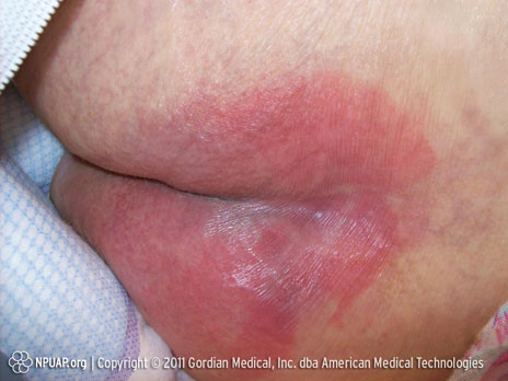
Pressure ulcer Category/Stage I: Non-blanchable redness of intact skin

Buttocks, Stage I, NPUAP copyright & used with permission.

Intact skin with non-blanchable erythema of a localised area, usually over a bony prominence. Discoloration of the skin, warmth, oedema, hardness or pain may also be present. Darkly pigmented skin may not have visible blanching.


Category/Stage II: Partial thickness skin loss or blister
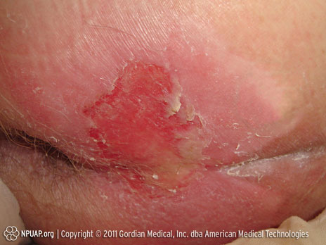
Pressure ulcer Category/Stage II: Partial thickness skin loss or blister

Buttocks, Stage II, NPUAP copyright & used with permission.
Partial thickness loss of dermis presenting as a shallow open ulcer with a red-pink wound bed, without slough. May also present as an intact or open/ruptured serum-filled or sero-sanginous-filled blister.


Category/Stage III: Full thickness skin loss (fat visible)
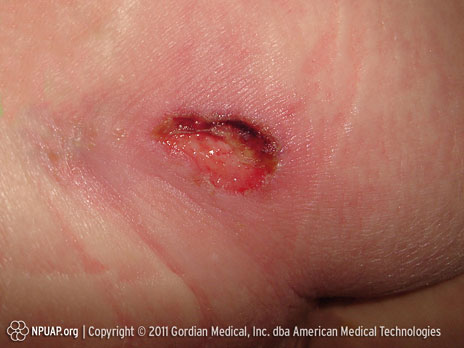
Pressure ulcer Category/Stage III: Full thickness skin loss (fat visible)

Ischium, Stage III, NPUAP copyright & used with permission.

Full thickness tissue loss. Subcutaneous fat may be visible, but no bone, tendon or muscle is exposed. Some slough may be present. May include undermining and tunnelling.


Category/Stage IV: Full thickness tissue loss (muscle/bone visible)
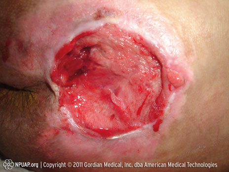
Pressure ulcer Category/Stage IV: Full thickness tissue loss (muscle/bone visible) 

Sacral Coccyx, Stage IV, NPUAP copyright & used with permission.

Full thickness tissue loss with exposed bone, tendon or muscle. Slough or eschar may be present. Often include undermining and tunnelling.


Treatment needs (2,3)

  • Pressure must be relieved or removed by appropriate measures.
  • Wound care must be optimised by:
  1. Debridement of necrotic tissue
  2. Appropriate cleansing of wound and surrounding skin
  3. Use of appropriate moist wound healing dressings

Suitable wound dressings for pressure injuries are foam or alginate dressings with superior absorption and exudate management properties, such as Biatain Silicone or Biatain Adhesive foam dressing or Biatain Alginate dressing. A silver-releasing dressing such as Biatain Ag can help prevent or resolve wound infection.

If the wound is painful, a moist wound healing foam dressing containing ibuprofen such as Biatain Ibu is a good option. The use of Biatain Ibu is supported by several randomised controlled studies. (4-6)


Download our tips for preventing, assessing and treating pressure ulcers:

Pressure injuries – prevention and treatment: A Coloplast quick guide (pdf)

Close

Acute wounds

Acute wounds must be treated promptly to stop bleeding and protect tissue. Read on to learn more about managing acute wounds to support faster healing

References

1. Enoch and Price 2004. (http://www.worldwidewounds.com/2004/august/Enoch/Pathophysiology-Of-Healing.html)

2.Winter. Nature  1962;193:293

3. Winter. Journal of Tissue Viability 2006;16(2):20-23

An acute wound is an injury that causes a break in the skin and sometimes the tissue. Acute wounds are classified into two principal types:

  • Acute traumatic wounds, such as abrasions, lacerations, penetrations or bites, and burns
  • Acute surgical wounds resulting from surgical incisions

acute wound


Acute wounds – signs and symptoms

  • A cut or tear in the skin
  • Bleeding, swelling, pain, and/or difficulty moving the affected area
  • There may be dirt or foreign objects inside the wound
  • Exudate is normally clear

Treatment needs

Large or deep, acute wounds with heavy bleeding need acute medical attention to stop bleeding and check for damage to vital organs or tissue.

Foreign objects should be removed from the wound and necrotic tissue debrided, as it can function as a base for infection and delay wound healing. Wound cleansing with clean water or antiseptics can be helpful.

Exudate production is part of the natural wound healing process (1), but the exudate needs to be managed appropriately. Exudate levels are often high during the inflammatory phase of wound healing, and leakage of exudate under the dressing can damage the surrounding skin. Wound exudate must be absorbed and managed by a dressing with moist wound healing properties. This supports the healing process and reduces scar formation. (2-3)

With appropriate care, smaller acute wounds will normally close within days or weeks depending on the size, depth and position of the wound.

Suitable dressings for acute wounds are dressings with superior absorption and exudate management properties that support a moist wound healing environment. These include Biatain Silicone, Biatain Adhesive or Comfeel. If the wound is painful, Biatain Ibu can be used. Where infection is present, Biatain Ag is an appropriate choice.


Why do some acute wounds become chronic?

If a person has a condition affecting the circulation system and/or immune response, normal wound healing processes may be impaired, and the wound may become chronic. Chronic wounds are often defined by the underlying condition that prevents wound healing, such as venous leg ulcers, arterial leg ulcers, diabetic foot ulcers or pressure injuries.

Close

Wound and skin condition

Necrosis

If a wound contains dead (necrotic) tissue, it’s a sign that the wound is not healing normally. Learn more about debridement of necrotic tissue

Necrotic tissue is often black or yellow. It may be soft or it may form a scab (eschar). Necrotic tissue can contain bacteria – and if the bacteria grow, the wound will become infected.

Necrosis


Removal of necrotic tissue (debridement)

Necrotic tissue must be removed in order to support wound healing. Removal can be surgical, mechanical, enzymatic (such as maggot therapy), or it can be achieved by supplementing the body’s own ability to break down necrotic tissue (autolytic debridement). 


Choice of dressing

The autolytic debridement process is optimised when the wound is moist. Coloplast has a range of dressings that support moist wound healing. For optimal moisture balance in the wound, the hydrocolloid Comfeel dressing and the unique properties of the Biatain foam dressing both support the natural autolytic debridement process.


Purilon gel is ideal for gentle and effective autolytic debridement of tissue in both dry and moist necrotic wounds, when used in combination with a secondary dressing such as Comfeel or Biatain.

Close

Peri-ulcer skin

The skin surrounding an ulcer is vulnerable. This may be associated with age, disease processes or exposure of the skin to wound exudate or dressing adhesives. Learn more about peri-ulcer skin.

Maceration 

Exudate leaked from ulcers can cause maceration, a softening or sogginess and breakdown of the skin that results from on-going contact with excessive moisture. Maceration can lead to skin breakdown, causing the ulcer to grow or creating satellite ulcers. Macerated tissue is white in colour.

macerated skin


Erythema

Erythema is an abnormal redness of the skin caused by dilation of blood vessels. Redness of the peri-ulcer skin may be a sign of inflammation or wound infection. 


Fragile skin

As we grow older, the texture of our skin changes and our skin becomes thinner, weaker and less protective. If a wound is surrounded by fragile skin, dressings are more likely to cause skin irritation. You must examine the skin carefully before deciding to use either an adhesive or a non-adhesive dressing.

Fragile skin


Choice of dressing

The Biatain dressing range offers dressings with superior absorption and exudate management properties for all types of skin conditions.

If a wound is surrounded by fragile skin, a dressing with silicone adhesive such as Biatain Silicone is an excellent alternative, as it can be used on both fragile and healthy skin.

Normal, healthy skin has a smooth and resilient structure. With proper wound treatment and use of dressings with superior absorption and exudate management, the skin surrounding a wound may be perfectly healthy and suitable for adhesive dressings such as Biatain Adhesive or Biatain Super Adhesive.


If the skin is very fragile, consider using a non-adhesive dressing such as Biatain Non-adhesive or Biatain Alginate.

Close

Wound exudate

Whether a wound is low-, medium- or high-exuding, superior absorption and control of exudate are essential for optimal healing. Learn more about wound exudate

References

1. White RJ and Cutting KF. British Journal of Nursing 2003;12(20):1186-1201

2. Adderly UJ. Wound Care, March 2010:15-20

3. Colwell JC et al. Wound Ostomy Continence Nursing 2011;38(5):541-53

4. Enoch B and Harding K. Wounds: A Compendium of Clinical Research and Practice 2003;15(7):213-29

5. Andersen et al. Ostomy/Wound Management 2002;(48)8:34-41

6. Thomas et al. http://www.dressings.org/TechnicalPublications/PDF/Coloplast-Dressings-Testing-2003-2004.pdf

7. White R and Cutting KF. http://www.worldwidewounds.com/2006/september/White/Modern-Exudate-Mgt.html

8. Romanelli et al. Exudate management made easy. Wounds International 2010;1(2).

In the inflammatory phase of wound healing, exudate levels are usually high. Non-healing, or chronic, wounds are often stuck in the inflammatory phase and may produce large amounts of exudate. Increased exudate levels can be a symptom of infection and increased oedema.

Wound exudate is a fluid composed of plasma, blood cells and platelets. Most of the wound exudate filters from the blood and/or lymph system into the wound area, but red blood cells and platelets leak from injured capillaries. Composition and viscosity varies, from thin and clear plasma fluid to thick yellow secretion containing high concentrations of white blood cells and bacteria.


Controlling exudate

If wound exudate is not properly controlled, it can leak from the dressing and result in the peri-ulcer skin being exposed to the exudate (1). This causes over-hydration maceration of the skin and can ultimately delay healing (2,3).

Maceration is a softening or sogginess and breakdown of the skin caused by on-going contact with excessive moisture. Macerated tissue looks white and maceration can cause an ulcer to grow or create satellite ulcers.


Macerated skin(2,3)

  • Delays healing
  • Increases risk of infection
  • Increases friction risk
  • Can result in wound enlargement

macerated skin


It is therefore very important that excess exudate is removed from the wound by an absorbent dressing. (4)

Control of exudate, removal of unhealthy tissue by debridement and management of bacterial load are all part of good wound bed preparation. The optimal wound dressing keeps the wound moist and absorbs exudate, locking it inside the dressing to prevent maceration.


Dressings for exuding wounds

We recommend the Biatain dressing range, which provides superior absorption - faster healing (5,6) of low to high exuding wounds. Biatain dressings effectively absorb and retain wound exudate, ensuring  a moisture balance that is optimal for healing of exuding wounds.(7,8)

Close

Wound infection

All wounds contain bacteria – even wounds that are healing normally. But if the bacteria count rises the wound may become infected. Learn more about treating infected wounds

References

1. Jørgensen et al. International Wound Journal 2005;2(1):64-73

2. Münter et al. Journal of Wound Care 2006;15(5):199-206

3. Reitzel & Marburger. EWMA 2009

4. Ip et al. Antimicrobial activities of silver dressings: an in vitro comparison. Journal of Medical Microbiology 2006;(55):59-63.

5. Basterzi et al. In-vitro comparison of antimicrobial efficacy of various wound dressings. Wounds 2010; July.

6. Data on file (Independent laboratory testing performed by Wickham Laboratories).

7. Thomas et al. www.dressings.org/TechnicalPublications/PDF/ Coloplast-Dressings-Testing-2003-2004.pdf

Wounds that are not healing normally may have a bacterial imbalance resulting in local infection of the wound.

Likely signs of wound infection are one or more of the following:

  • Delayed or stalled healing
  • Odour
  • Increased wound exudate
  • Absent/abnormal/discoloured granulation tissue
  • Increased pain at wound site

infected wound


Other symptoms

Additional clinical symptoms may arise if the infection spreads to the healthy tissue surrounding the wound. Depending on the type of bacteria, the wound exudate may become more pus-like, and the peri-ulcer skin may be tender, red and painful. The patient may also have a fever.

Please remember that diabetic foot ulcers do not always present with the classical signs of local infection.


Dressings for infected wounds and wounds at risk of infection

If a wound is healing normally, a foam dressing with superior absorption such as Biatain or Biatain Silicone is ideal. If the wound is infected or there is risk of wound infection, we recommend a silver dressings such as Biatain Ag. This provides superior absorption for faster healing of infected wounds (1-7). If the infection is spreading beyond the wound, the silver dressing must be combined with systemic antibiotic treatment at the discretion of a physician. 

Close

Wound pain

Studies show that more than 80% of patients with chronic wounds are in constant pain and half of them classify the pain as moderate to severe. (1-6) Wound pain and local pain management options

References

1. Gottrup et al. Wound Repair and Regeneration 2008;16:616-26

2. Fogh et al. Wound Repair and Regeneration 2012;20:815-21

3. Palao I Domenech et al. Journal of Wound Care 2008;17(8):342-48

4. Woo et al. International Wound Journal 2008;5(2):205-215

5. Pieper et al. Ostomy Wound Management 1998;44:54–58&60–67

6. Nemeth et al. Ostomy Wound Management 2004;50:34–36

Many patients say that the pain is the worst aspect of having a wound. Persistent or chronic wound pain not only affects patients’ quality of life, it can also be a major barrier to wound healing. Often, the consequences of painful wounds are underestimated and undertreated. 
The wound pain itself can be caused by tissue damage (nociceptive) or nerve damage (neuropathic).

Nociceptive pain: stimulus dependent and usually caused by tissue damage. The pain is described as gnawing, aching, throbbing and tender. 

Neuropathic pain: occurs spontaneously as a result of nerve tissue injury. The pain is most likely to be described as burning, stinging, shooting or stabbing. 


Management of wound pain 

First, treat the possible causes of pain, such as wound infection (e.g., by a silver dressing) and uncontrolled oedema.

Local pain management is an important first step for persistent wound pain and for painful procedures, such as dressing changes and debridement.

You should consider systemic treatment if the pain does not improve with local pain management.

painful wound


Biatain Ibu – Superior absorption for painful wounds

Biatain Ibu is the first and only dressing to combine moist wound healing with local release of ibuprofen (1,2). Biatain Ibu is indicated for exuding wounds to provide moist wound healing. In addition, Biatain Ibu may reduce wound pain caused by tissue damage (nociceptive pain). The safety and performance of Biatain Ibu is supported by evidence from the largest randomised, comparative clinical trials ever performed on a wound dressing(1-3).

Close

Case studies

Treatment of a venous leg ulcer

Treatment of a venous leg ulcer

See how the use of Biatain® Non-Adhesive foam dressings led to a 95% reduction in ulcer area after four weeks’ treatment of a venous leg ulcer. Read more

The patient

The patient – an 85-year-old woman – had been suffering from a venous leg ulcer on the lateral part of her lower left leg. The skin on her leg was fragile. The ulcer had persisted for five months at inclusion.


Previous treatment

Prior to inclusion, the ulcer had been treated with alginate dressings and compression therapy for ten weeks. Ulcer healing was delayed compared to the normally expected healing rate.


Biatain Non-Adhesive introduced

 When the patient began treatment with Biatain Non-Adhesive, the ulcer area was 4.9 cm2. The ulcer contained 20% fibrinous tissue and 80% healthy granulation tissue. During the four-week treatment period, long-stretch compression bandages were applied.

This picture shows the ulcer at inclusion after cleansing:
The ulcer at inclusion after cleansing.

Ulcer area was reduced by 73% after two weeks of treatment with Biatain Non-Adhesive:
Ulcer area was reduced by 73% after two weeks treatment.

The ulcer area was reduced by 95% after 4 weeks of treatment with Biatain Non-Adhesive:
After four weeks treatment the ulcer area was reduced by 95%.


Conclusion

During the four weeks treatment period:

  • Biatain Non-Adhesive demonstrated remarkable reduction in ulcer size.
  • Biatain Non-Adhesive caused no leakage or maceration, even under compression therapy.
  • Biatain Non-Adhesive minimised pressure marks.
  • Biatain Non-Adhesive was comfortable and easy to use.


Download full case description (PDF)

Close
Treatment of an infected diabetic foot ulcer

Treatment of an infected diabetic foot ulcer

See how treatment of a heavily infected ulcer with Biatain® Ag Non-Adhesive foam dressing and Biatain Alginate® Ag prevented amputation of a foot. Read more

The patient

This patient suffered from heart failure, coronary heart disease, arterial hypertension and venous insufficiency in both lower limbs, as well as Alzheimer’s.


Deep foot ulcer

For four months, she had a very deep diabetic foot ulcer on the right foot with erythema, oedema, crepitation and heat in the surrounding tissues. She was referred to the A&E department and after examination by Vascular Surgery, her family was informed that the immediate treatment would involved supracondylar amputation since she was suffering from a grade 4–5 diabetic foot based on the Wagner scale.
 

Biatain Ag introduced

The family was opposed to this treatment, and the patient was returned to her home for monitoring by her family doctor and out-patient care and dressing by home nursing. Biatain Alginate Ag and Biatain Ag were used in combination with debridement and oral antibiotics.

This picture shows the ulcer at inclusion:
The ulcer at inclusion.

This picture shows the ulcer after five weeks of treatment with first Biatain Alginate Ag and then Biatain Ag Non-Adhesive:
The ulcer after 5 weeks treatment with first seasorb Ag and then Biatain Ag.

Here we see the ulcer after four months’ treatment:
The ulcer after 4 months treatment.

The ulcer was closed after 10 months’ treatment:
Closed ulcer after 10 months treatment


Conclusion

The objective set was to prevent amputation of the foot, ensure the well being and comfort of the patient and of the family. The method used began to bear fruit already seven days after the treatment started, with visible changes in the development of the wound. The wound closed ten months after the treatment was started.

Download full case description (PDF)

Tips for prevention, assessment and treatment of diabetic foot ulcers are available in: Diabetic foot ulcers – prevention and treatment: A Coloplast quick guide.

Close
Treatment of a Stage III sacral pressure injury

Treatment of a Stage III sacral pressure injury

See how a one-month treatment of a heavily exuding sacral pressure ulcer with Biatain® Ag effectively eliminated signs of local infection. Read more

The patient

The patient was an 88-year-old woman with a highly exuding Stage III sacral pressure injury. The ulcer had persisted for two months and had previously been treated with standard moist wound healing products.
 

The ulcer

The ulcer had several signs of local infection, a significant odour, and was heavily exuding. A small undermining was present at the top of the ulcer and there was approximately 50% unhealthy necrotic tissue in the wound bed.

The first picture shows the infected, highly exuding pressure injury before Biatain Ag treatment:

The ulcer at the start of treatment.

Biatain Ag Adhesive introduced

Odour was eliminated after just one week of treatment:

The ulcer after one week of treatment.

This picture shows that the wound bed is clean and healing is progressing after four weeks of treatment:

The ulcer after four weeks of treatment.


Conclusion

During the one-month treatment period Biatain Ag effectively eliminated signs of local infection and supported healing of this heavily exuding sacral pressure injury. Elimination of odour and a significant increase in healthy granulation tissue was observed already after one week.


Download full case description (PDF)

Tips for prevention, assessment and treatment of pressure injuries are available in:

Pressure injuries – prevention and treatment: A Coloplast quick guide

Close
Close

Free sample - Free shipping

Thank you for your order

Confirmation

View desktop version